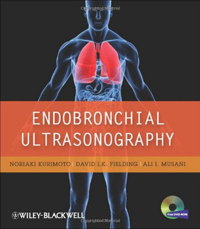
Endobronchial Ultrasonography 2nd Edition by Noriaki Kurimoto, David Fielding, Ali Musani, Christopher Kniese, Katsuhiko Morita ISBN 111923395X 9781119233954
$70.00 Original price was: $70.00.$35.00Current price is: $35.00.
Instant download Endobronchial Ultrasonography Kurimoto Noriaki. Fielding David I. K. Musani Ali I after payment
Endobronchial Ultrasonography 2nd Edition by Noriaki Kurimoto, David I. K. Fielding, Ali I. Musani, Christopher Kniese, Katsuhiko Morita – Ebook PDF Instant Download/Delivery: 111923395X, 9781119233954
Full dowload Endobronchial Ultrasonography 2nd Edition after payment

Product details:
ISBN 10: 111923395X
ISBN 13: 9781119233954
Author: Noriaki Kurimoto, David I. K. Fielding, Ali I. Musani, Christopher Kniese, Katsuhiko Morita
Endobronchial ultrasonography (EBUS) is an exciting and still developing diagnostic tool that has added significantly to the diagnosis and staging of lung cancer and other thoracic diseases. Co-authored by one of the technology’s pioneers, this book helps the reader to use EBUS to diagnose and stage lung cancer and a variety of different tumours of the chest region. The second edition of Endobronchial Ultrasonography covers all of the standard techniques and the very latest developments and guidelines involved in EBUS, combining two common procedures, bronchoscopy and real-time ultrasonography, allowing physicians to obtain precise biopsies of lymph nodes and masses within the chest cavity.
Endobronchial Ultrasonography 2nd Table of contents:
1 Endobronchial Ultrasonography: An Overview
Introduction
Principles of Ultrasonography
Equipment
Preparations
Operation
Equipment for EBUS‐Guided TBNA
References
2 Identifying Peribronchial Organs during Endobronchial Ultrasonography
Overview of Ultrasound Imaging of the Bronchi as the Radial Probe Is Pulled in a Proximal Direction
Ultrasound Imaging of Mediastinal and Hilar Lymph Nodes for EBUS‐TBNA Using a Convex Bronchoscope
Azygous Vein and Esophagus
Reference
3 How to Perform Endobronchial Ultrasonography
Introduction
Balloon Probes for Central Lesions
Performing EBUS Using a Guide Sheath for Examining Peripheral Pulmonary Lesions
EBUS‐TBNA
New Functions of Ultrasound Processors
References
4 Endobronchial Ultrasound‐Guided Transbronchial Needle Aspiration
Anatomy
Transbronchial Needle Aspiration (TBNA)
Needles
Planning the Procedure
Insertion Technique
Sample Handling
Endobronchial Ultrasound
Step‐by‐Step EBUS‐TBNA
Acknowledgments
References
5 Endobronchial Ultrasound‐Guided Transbronchial Needle Aspiration: Tips and Tricks
Choice of Sedation Type
Considerations Regarding Presumed Tissue Type
Other Causes of PET‐Positive Nodes with Negative EBUS‐TBNA (False‐Positive PET)
Technical Aspects Including Balloons, Stylet, Use of the Sheath
Diagnosing Primary Masses
Genetics on EBUS‐TBNA Lung Cancer Samples
Next‐Generation Sequencing (NGS)
EBUS‐TBNA and PD‐L1 Staining
References
6 Endoscopic Ultrasound‐Guided Mediastinal Lymph Node Aspiration for Lung Cancer Diagnosis and Staging
Introduction
Technique
Role of EUS in Lung Cancer Staging
Combination of EUS and EBUS for Mediastinal Lymph Node Sampling
References
7 How to Accurately Identify the Bronchial Pathway to a Peripheral Pulmonary Lesion
Introduction
Reading CT Anatomy versus Virtual Bronchoscopic Navigation
Step‐by‐Step Guide to Reading CT Anatomy
Representative Cases
Future Directions
8 Qualitative Analysis of Peripheral Pulmonary Lesions Using Endobronchial Ultrasonography
Introduction
Internal Structure of Peripheral Pulmonary Lesions Visualized by EBUS
Internal Structural Components in EBUS Images of Peripheral Pulmonary Lesions
Classifications of Lesions Based on Internal Structures Visualized by EBUS
Articles and Discussion
References
9 Diagnosis of Peripheral Pulmonary Lesions Using Endobronchial Ultrasonography with a Guide Sheath
Introduction
Equipment
EBUS‐GS Procedure
Effectiveness of Using EBUS‐GS to Diagnose PPLs
Changes in EBUS‐GS Techniques
How to Identify the Drainage Bronchus Leading to the Target Lesion
Signal Attenuation Caused by the Guide Sheath
Moving the Probe within the Lesion
Management of Post‐Biopsy Hemorrhaging
Conclusion
References
10 Endobronchial Ultrasonography with a Guide Sheath: Up to Date
Introduction
Literature Review Including Meta‐analyses
Up‐to‐Date Advances in EBUS‐Guided Peripheral Nodule Biopsy
References
11 Endobronchial Ultrasonography for Ground‐Glass Opacity Lesions
References
12 Techniques for Comparing Endobronchial Ultrasonography Images of Peripheral Pulmonary Lesions with Macroscopy and Histopathology Findings
Introduction
“Branch Reading”: Identification of Involved Bronchi Leading to Target Lesion Using CT Images
Diagnostic Bronchoscopy
Bronchoscopy of the Excised Specimen
Endobronchial Ultrasonography: Case Presentation
13 Endobronchial Ultrasonography of Airway Integrity and Tumor Involvement
Introduction
Optical Coherence Tomography
References
14 Endobronchial Ultrasonography in Interventional Bronchoscopy
Introduction
Technique
Evaluating Tracheal Compression versus Tracheal Wall Invasion
Relapsing Polychondritis with Tracheobronchomalacia
Endobronchial Stenting
Brachytherapy
Thermal Applications
Miscellaneous Applications
References
15 Cytopathology in Endobronchial Ultrasound‐Guided Transbronchial Needle Aspiration of Mediastinal and Hilar Lymph Nodes
Introduction
Technique
Role of Rapid On‐Site Evaluation in Endobronchial Ultrasonography
Recent Advances in ROSE during EBUS‐TBNA
References
16 Future Directions for Endobronchial Ultrasonography
Peripheral Pulmonary Lesions and EBUS
Other Technical Innovations Combined with EBUS GS
People also search for Endobronchial Ultrasonography 2nd:
endobronchial ultrasonography
bronchoscopy and endobronchial ultrasonography
endobronchial ultrasonography lung cancer
endobronchial ultrasonography definition
endobronchial ultrasonography wiki


