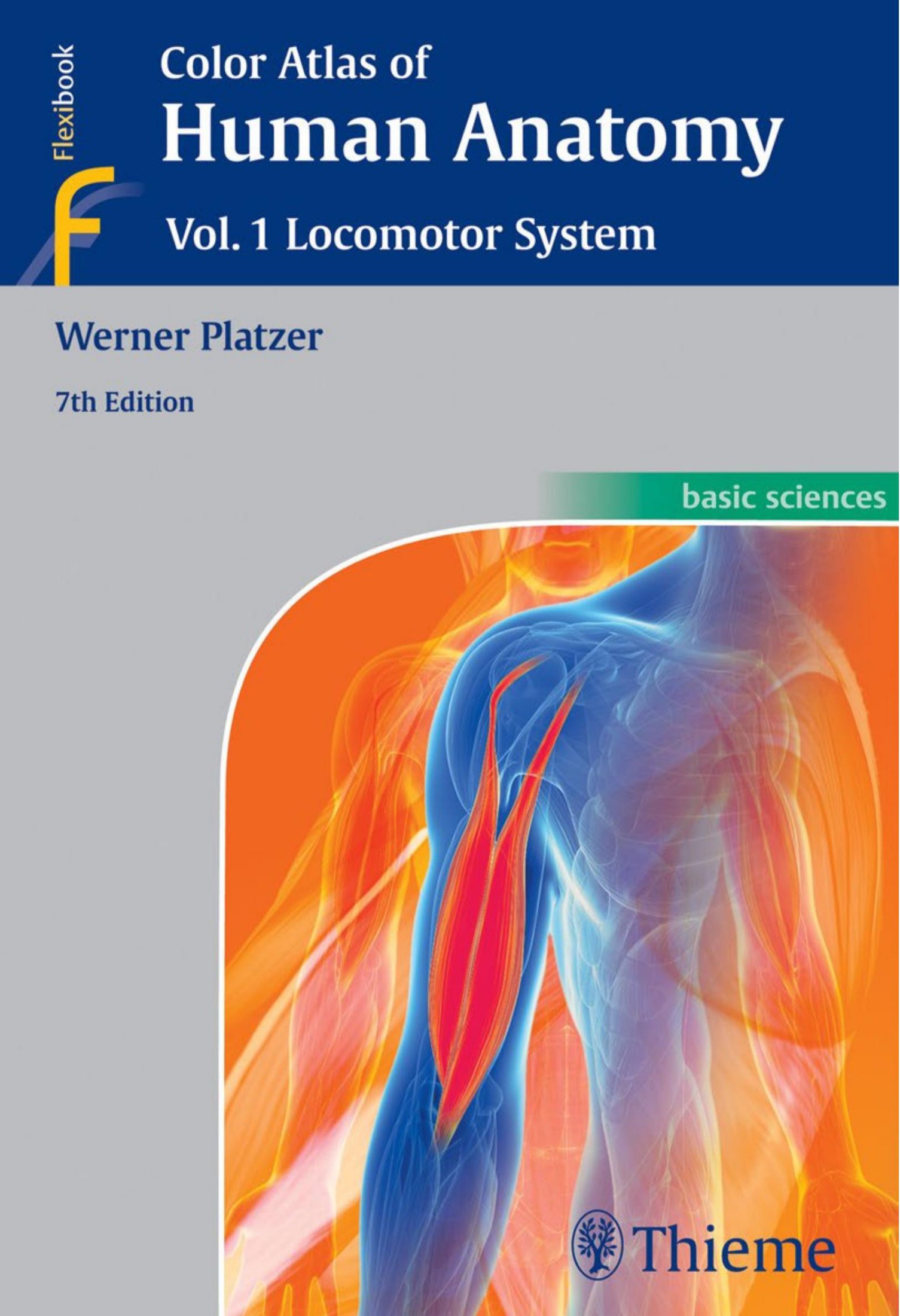
Color Atlas of Human Anatomy Volume 1 Locomotor System 7th edition by Werner Platzer ISBN 3135333078 978-3135333076
$70.00 Original price was: $70.00.$35.00Current price is: $35.00.
Instant download Color Atlas of Human Anatomy 7e Volume 1 Locomotor System Wei Zhi after payment
Color Atlas of Human Anatomy Volume 1 Locomotor System 7th edition by Werner Platzer- Ebook PDF Instant Download/Delivery: 3135333078 978-3135333076
Full download Color Atlas of Human Anatomy Volume 1 Locomotor System 7th edition after payment

Product details:
ISBN 10: 3135333078
ISBN 13: 978-3135333076
Author: Werner Platzer
The seventh edition of this classic work makes mastering large amounts of complex information much less daunting. These are some of the many user-friendly features of this book:
- More than 200 outstanding full-color illustrations and 100 new clinical correlations
- Side-by-side images with updated callouts
- An overview of anatomical terms and their Latin equivalents in each section
Emphasizing clinical anatomy, this atlas integrates current information from a wide range of medical disciplines into the discussions of the locomotor system, including:
- General anatomy
- The systematic anatomy of the locomotor system
- The topography of peripheral nerves and vessels in relation to the musculoskeletal system
Volume 1: Locomotor System and its companions Volume 2: Internal Organs and Volume 3: Nervous System and Sensory Organs comprise a must-have resource for students of medicine, dentistry, and all allied health fields.
Color Atlas of Human Anatomy Volume 1 Locomotor System 7th Table of contents:
- Preface
- General Anatomy
- The Body
- Parts of the Body
- General Terms
- The Cell
- Cytoplasm
- Cell Nucleus
- Vital Cell Functions
- Tissues
- Epithelia
- Connective Tissue and Supporting Tissues
- Connective Tissue
- Cartilage
- Bone
- Development of Bone
- Muscular Tissue
- General Features of the Skeleton
- Classification of Bones
- Periosteum
- Joints between Bones
- Continuous Joints between Bones
- Discontinuous Joints between Bones
- Types of Discontinuous (Synovial) Joints
- General Features of the Muscles
- Classification of Skeletal Muscles
- Auxiliary Features of Muscles
- Investigation of Muscle Function
- Anatomical Terms and their Latin Equivalents
- Systematic Anatomy of the Locomotor System
- Trunk
- Vertebral Column
- Cervical Vertebrae
- Thoracic Vertebrae
- Lumbar Vertebrae
- Malformations and Variations of the Presacral Vertebral Column
- Sacrum
- Coccyx
- Variations in the Sacral Region
- Ossification of the Vertebrae
- Intervertebral Disks
- Ligaments of the Vertebral Column
- Joints of the Vertebral Column
- The Vertebral Column as a Whole
- Thoracic Cage
- Ribs
- Sternum
- Joints of the Ribs
- Structure of the Thoracic Cage
- Movements of the Thoracic Cage
- Intrinsic Muscles of the Back
- Suboccipital Muscles
- Body Wall
- Thoracolumbar Fascia
- Extrinsic Ventrolateral Muscles
- Prevertebral Muscles
- Scalene Muscles
- Muscles of the Thoracic Cage
- Intercostal Muscles
- Abdominal Wall
- Superficial Abdominal Muscles
- Function of the Superficial Abdominal Musculature
- Fascias of Abdominal Wall
- Deep Abdominal Muscles
- Sites of Weakness in the Abdominal Wall
- Diaphragm
- Position and Function of the Diaphragm
- Sites of Diaphragmatic Hernias
- Pelvic Floor
- Pelvic Diaphragm
- Urogenital Diaphragm
- Anatomical Terms and their Latin Equivalents
- Upper Limb
- Bones, Ligaments and Joints
- Shoulder Girdle
- Scapula
- Ligaments of the Scapula
- Clavicle
- Joints of the Shoulder Girdle
- The Free Upper Limb
- Bone of the Arm
- Shoulder Joint
- Movements of the Shoulder Joint
- Bones of the Forearm
- Elbow Joint
- Distal Radioulnar Joint
- Continuous Fibrous Joint between Radius and Ulna
- Carpus
- Individual Bones of the Carpus
- Bones of the Metacarpus and Digits
- Radiocarpal and Midcarpal Joints
- Movements in the Radiocarpal and Midcarpal Joints
- Carpometacarpal and Intermetacarpal joints
- Metacarpophalangeal and Digital Joints
- Muscles, Fascias, and Special Features
- Muscles of the Shoulder Girdle and Arm
- Classification of the Muscles
- Shoulder Muscles Inserted on the Humerus
- Trunk Muscles Inserted on the Shoulder Girdle
- Cranial Muscles Inserted on the Shoulder Girdle
- Function of the Shoulder Girdle Muscles
- Fascias and Spaces in the Shoulder Girdle Region
- Arm Muscles
- Muscles of the Forearm
- Classification of the Muscles
- Ventral Forearm Muscles
- Radial Forearm Muscles
- Dorsal Forearm Muscles
- Function of Muscles of the Elbow Joint and Forearm
- Function of Muscles of the Wrist and the Midcarpal Joint
- Intrinsic Muscles of the Hand
- Central muscles of the hand
- Thenar Muscles
- Palmar Aponeurosis
- Hypothenar Muscles
- Fascias and Special Features of the Free Upper Limb
- Fascias
- Tendinous Sheaths
- Anatomical Terms and their Latin Equivalents
- Lower Limb
- Bones, Ligaments, Joints
- Pelvis
- Hip Bone
- Junctions between the Bones of the Pelvis
- Morphology of the Bony Pelvis
- The Free Lower Limb
- Femur
- Patella
- Positions of the Femur
- Hip Joint
- Bones of the Leg
- Knee Joint
- Movements of the Knee Joint
- Alignment of the Lower Limb
- Connections between the Tibia and the Fibula
- Bones of the Foot
- Sesamoid Bones
- Joints of the Foot
- Ligaments of the Joints of the Foot
- Morphology and Function of the Skeleton of the Foot
- The Plantar Arch and Its Function
- Foot Types
- Muscles, Fascias, and Special Features
- Muscles of the Hip and Thigh
- Classification of the Muscles
- Dorsal Hip Muscles
- Ventral Hip Muscles
- Adductors of the Thigh
- Function of the Hip Muscles and Adductors of Thigh
- Anterior Thigh Muscles
- Posterior Thigh Muscles
- Function of the Knee Joint Muscles
- Fascias of the Hip and Thigh
- Long Muscles of the Leg and Foot
- Classification of the Muscles
- Anterior Leg Muscles
- Posterior Leg Muscles
- Function of the Ankle, Subtalar and Talocalcaneonavicular Joint Muscles
- Intrinsic Muscles of the Foot
- Muscles of the Dorsum of the Foot
- Muscles of the Sole of the Foot
- Fascias of the Leg and the Foot
- Tendinous Sheaths in the Foot
- Anatomical Terms and their Latin Equivalents
- Head and Neck
- Skull
- Subdivision of the Skull
- Ossification of the Skull
- Special Features of Intramembranous Ossification
- Sutures and Synchondroses
- Structure of the Cranial Bones
- Calvaria
- Lateral View of the Skull
- Posterior View of the Skull
- Anterior View of the Skull
- External surface of cranial base
- Internal surface of cranial base
- Variants of the Internal Surface of cranial base
- Sites for Passage for Vessels and Nerves
- Mandible
- Shape of Mandible
- Hyoid Bone
- Orbital Cavity
- Pterygopalatine Fossa
- Nasal Cavity
- Cranial Shapes
- Malformations
- Special Cranial Shapes and Sutures
- Accessory Bones of the Skull
- Temporomandibular Joint
- Muscles and Fascias
- Muscles of the Head
- Mimetic Muscles
- Muscles of Mastication
- Ventral Muscles of the Neck
- Infrahyoid Muscles
- Head Muscles Inserted on the Shoulder Girdle
- Fascias of the Neck
- Anatomical Terms and their Latin Equivalents
- Topography of Peripheral Nerves and Vessels
- Head and Neck
- Regions
- Anterior Facial Regions
- Orbital Region
- Lateral Facial Regions
- Infratemporal Fossa
- Superior View of the Orbit
- Occipital Region and Posterior Cervical (Nuchal) Region
- Suboccipital Triangle
- Lateral Pharyngeal and Retropharyngeal Spaces
- Submandibular Triangle
- Retromandibular Fossa
- Middle Region of the Neck
- Thyroid Region
- Ventrolateral Cervical Regions
- Scalenovertebral Triangle
- Upper Limb
- Regions
- Deltopectoral Triangle
- Axillary Region
- Axillary Spaces
- Anterior Region of Arm
- Posterior Region of Arm
- Cubital Fossa
- Anterior of forearm Region
- Anterior Region of wris
- Palm of Hand
- Dorsum of the Hand
- Radial Fovea, “Anatomical Snuff Box”
- Trunk
- Regions
- Regions of the Thorax
- Anterior Thorax Regions
- Posterior Thorax Regions
- Regions of the Abdomen
- Inguinal Region
- Lumbar Region
- Perineal Region of the Female
- Perineal Region of the Male
- Lower Limb
- Regions
- Subinguinal Region
- Saphenous Opening
- Gluteal Region
- Anterior Region of the Thigh
- Posterior Region of the Thigh
- Posterior Region of the Knee
- Popliteal Fossa
- Anterior Region of the Leg
- Posterior Region of the Leg
- Medial Retromalleolar Region
- Dorsum of the Foot
- Sole of the Foot
- Anatomical Terms and their Latin Equivalents
- For those who want to learn more
People also search for Color Atlas of Human Anatomy Volume 1 Locomotor System 7th:
color atlas of anatomy 8th edition
color atlas of human anatomy
color atlas of anatomy rohen pdf
a color atlas of parasitology sullivan pdf
a color atlas of parasitology pdf
Tags:
Werner Platzer,Color Atlas,Human Anatomy


