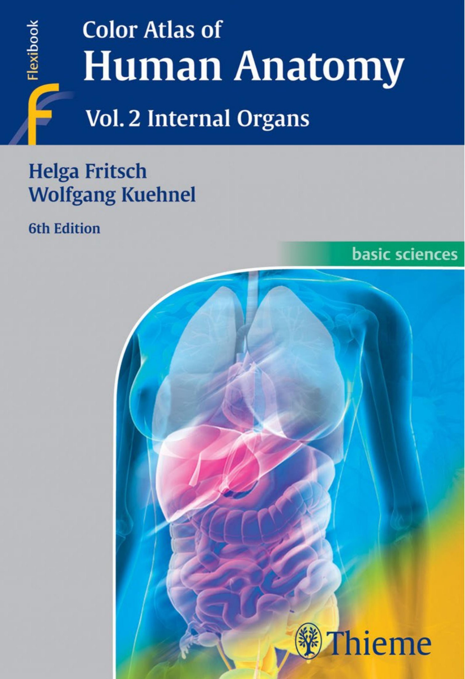
Color Atlas of Human Anatomy Volume 2 Internal Organs 6th edition by Helga Fritsch, Wolfgang Kuehnel ISBN 3135334066 978-3135334066
$70.00 Original price was: $70.00.$35.00Current price is: $35.00.
Instant download Color Atlas of Human Anatomy Volume 2 Internal Organs 6e Wei Zhi after payment
Color Atlas of Human Anatomy Volume 2 Internal Organs 6th edition by Helga Fritsch, Wolfgang Kuehnel – Ebook PDF Instant Download/Delivery: 3135334066 978-3135334066
Full download Color Atlas of Human Anatomy Volume 2 Internal Organs 6th edition after payment

Product details:
ISBN 10: 3135334066
ISBN 13: 978-3135334066
Author: Helga Fritsch, Wolfgang Kuehnel
The sixth edition of this classic work makes mastering a vast amount of information on internal organs much less daunting. It offers a vivid review of the human body and its structure, and it is an ideal study companion as well as an excellent basic reference text.
These are some of the many user-friendly features of this book
- New color plates on embryology and histology
- More than 200 outstanding full-color illustrations and 130 clinical correlations
- Side-by-side images with explanatory text
- An overview of anatomical terms in each section
Emphasizing clinical anatomy, this text integrates current information from a wide range of medical disciplines into discussions of the internal organs, including:
- Cross-sectional anatomy as a basis for working with modern imaging modalities
- Detailed explanations of organ topography and function
- Physiological and biochemical information included where appropriate
- An entire chapter devoted to pregnancy and human development
Volume 2: Internal Organs and its companions Volume 1: Locomotor System and Volume 3: Nervous System and Sensory Organs comprise a must-have resource for students of medicine, dentistry, and all allied health fields.
Color Atlas of Human Anatomy Volume 2 Internal Organs 6th Table of contents:
- Preface
- Viscera at a Glance
- Arrangement by Function
- Arrangement by Region
- Cardiovascular System
- Overview
- Circulatory System and Lymphatic Vessels
- Fetal Circulation (A)
- Circulatory Adjustments at Birth (B)
- Heart
- External Features
- Chambers of the Heart
- Cardiac Skeleton
- Layers of the Heart Wall
- Layers of the Heart Wall, Histology, and Ultrastructure
- Heart Valves
- Vasculature of the Heart
- Conducting System of the Heart
- Innervation
- Pericardium
- Position of the Heart and Cardiac Borders
- Radiographic Anatomy
- Auscultation
- Cross-Sectional Anatomy
- Cross-Sectional Echocardiography
- Functions of the Heart
- Arterial System
- Aorta
- Arteries of the Head and Neck
- Common Carotid Artery
- External Carotid Artery
- Maxillary Artery
- Internal Carotid Artery
- Subclavian Artery
- Arteries of the Shoulder and Upper Limb
- Axillary Artery
- Brachial Artery
- Radial Artery
- Ulnar Artery
- Arteries of the Pelvis and Lower Limb
- Internal Iliac Artery
- External Iliac Artery
- Femoral Artery
- Popliteal Artery
- Arteries of the Leg and Foot
- Vascular Arches of the Feet
- Venous System
- Caval System
- Azygos Vein System
- Tributaries of the Superior Vena Cava
- Brachiocephalic Veins
- Jugular Veins
- Dural Venous Sinuses
- Veins of the Upper Limb
- Tributaries of the Inferior Vena Cava
- Iliac Veins
- Veins of the Lower Limb
- Lymphatic System
- Lymphatic Vessels
- Regional Lymph Nodes of the Head, Neck, and Arm
- Regional Lymph Nodes of the Thorax and Abdomen
- Regional Lymph Nodes of the Pelvis and Lower Limb
- Structure and Function of Blood and Lymphatic Vessels
- Vessel Wall
- Special Forms of Arteries
- Regional Variation in Vessel Wall Structure—Arterial Vessels
- Regional Variation in Vessel Wall Structure—Venous Vessels
- Respiratory System
- Overview
- Anatomical Division of the Respiratory System
- Clinical Division of the Respiratory System
- Nose
- External Nose
- Nasal Cavity
- Paranasal Sinuses
- Openings of Paranasal Sinuses and Nasal Meatuses
- Posterior Nasal Apertures
- Nasopharynx
- Larynx
- Laryngeal Skeleton
- Structures Connecting the Laryngeal Cartilages
- Laryngeal Muscles
- Laryngeal Cavity
- Glottis
- Trachea
- Trachea and Extrapulmonary Main Bronchi
- Topography of the Trachea and Larynx
- Lung
- Surfaces of the Lung
- Divisions of the Bronchi and Bronchopulmonary Segments
- Microscopic Anatomy
- Vascular System and Innervation
- Pleura
- Cross-Sectional Anatomy
- Mechanics of Breathing
- Mediastinum
- Right View of the Mediastinum
- Left View of the Mediastinum
- Alimentary System
- Overview
- General Structure and Functions
- Oral Cavity
- General Structure
- Palate
- Tongue
- Muscles of the Tongue
- Inferior Surface of the Tongue (A)
- Floor of the Mouth
- Salivary Glands
- Microscopic Anatomy of the Salivary Glands
- Teeth
- Parts of the Tooth and the Periodontium
- Deciduous Teeth
- Eruption of the Primary and Permanent Dentition
- Development of the Teeth
- Position of the Teeth in the Dental Arcades
- Pharynx
- Organization and General Structure
- The Act of Swallowing
- Topographical Anatomy I
- Sectional Anatomy of the Head and Neck
- Esophagus
- General Organization and Microscopic Anatomy
- Topographical Anatomy of the Esophagus and the Posterior Mediastinum
- Vessels, Nerves, and Lymphatic Drainage
- Abdominal Cavity
- General Overview
- Topography of the Opened Abdominal Cavity
- Relations of the Parietal Peritoneum
- Stomach
- Gross Anatomy
- Microscopic Anatomy of the Stomach
- Vessels, Nerves, and Lymphatic Drainage
- Small Intestine
- Gross Anatomy
- Structure of the Small Intestinal Wall
- Vessels, Nerves, and Lymphatic Drainages
- Large Intestine
- Segments of the Large Intestine: Overview
- Cecum and Vermiform Appendix
- Colon Segments
- Rectum and Anal Canal
- Liver
- Gross Anatomy
- Liver Segments
- Microscopic Anatomy
- Portal Vein System (C)
- Bile Ducts and Gallbladder
- Gallbladder
- Pancreas
- Gross and Microscopic Anatomy
- Topography of the Omental Bursa and Pancreas
- Topographical Anatomy II
- Sectional Anatomy of the Upper Abdomen
- Sectional Anatomy of the Upper and Lower Abdomen
- Urinary System
- Overview
- Organization and Position of the Urinary Organs
- Kidney
- Gross Anatomy
- Microscopic Anatomy
- Topography of the Kidneys
- Excretory Organs
- Renal Pelvis and Ureter
- Urinary Bladder
- Female Urethra
- Topography of the Excretory Organs
- Male Genital System
- Overview
- Male Reproductive Organs
- Testis and Epididymis
- Gross Anatomy
- Microscopic Anatomy
- Seminal Ducts and Accessory Sex Glands
- Ductus Deferens (Vas Deferens)
- Seminal Vesicles
- Prostate
- Male External Genitalia
- Penis
- Male Urethra
- Topographical Anatomy
- Sectional Anatomy
- Female Genital System
- Overview
- Female Reproductive Organs
- Ovary and Uterine Tubes
- Gross Anatomy of the Ovary
- Microscopic Anatomy of the Ovary
- Follicles
- Gross Anatomy of the Uterine Tube
- Microscopic Anatomy of the Uterine Tube
- Uterus
- Gross Anatomy
- Microscopic Anatomy
- Vessels, Nerves, and Lymphatic Drainage
- Support of the Uterus
- Vagina and External Genitalia
- Gross Anatomy
- Topographical Anatomy
- Sectional Anatomy
- Comparative Anatomy of the Female and Male Pelves
- Soft Tissue Closure of the Pelvis
- Pregnancy and Human Development
- Gametes
- Fertilization
- Capacitation and Acrosome Reaction
- Formation of the Zygote
- Early Development
- Pregnancy
- Placenta
- Birth (Parturition)
- Dilation Stage
- Expulsion Stage
- Overview
- Prenatal Period
- Stages in Prenatal Development
- Pre-embryonic Period
- Embryonic Period
- Fetal Period (Overview)
- Fetal Period (Monthly Stages)
- Organ Development
- Body Cavities
- Heart
- Vessel Development
- Respiratory System
- Gastrointesinal System, Foregut
- Gastrointestinal System, Midgut and Hindgut
- Development of the Urinary System
- Development of the Genital System
- The Neonate
- Postnatal Periods
- Endocrine System
- Glands
- Overview
- Light Microscopic Classification of Exocrine Secretory Units
- General Principles of Endocrine Gland Function
- Hypothalamic–Pituitary Axis
- Gross Anatomy
- Microscopic Structure of the Pituitary Gland
- Hypothalamus–Pituitary Connections
- Efferent Connections of the Hypothalamus
- Hypothalamic–Posterior Pituitary Axis (A)
- Hypothalamic–Anterior Pituitary Axis (B)
- Pineal Gland
- Gross Anatomy
- Microscopic Anatomy
- Adrenal Glands
- Gross Anatomy
- Microscopic Anatomy of the Adrenal Cortex
- Microscopic Anatomy of the Adrenal Medulla
- Thyroid Gland
- Gross Anatomy
- Microscopic Anatomy
- Parathyroid Glands
- Pancreatic Islets
- Microscopic Anatomy
- Diffuse Endocrine System
- Testicular Endocrine Functions
- Ovarian Endocrine Functions
- Ovarian Cycle
- Endocrine Functions of the Placenta
- Cardiac Hormones—Atrial Natriuretic Peptides
- Diffuse Endocrine Cells in Various Organs
- Blood and Lymphatic Systems
- Blood
- Components of Blood
- Hematopoiesis
- Immune System
- Cells of the Immune System
- Lymphatic Organs
- Overview
- Thymus
- Microanatomy of the Thymus
- Lymph Nodes
- Spleen
- Microscopic Anatomy of the Spleen
- The Tonsils
- Mucosa-Associated Lymphoid Tissue (MALT)
- The Integument
- Skin
- General Structure and Functions
- Skin Color
- Surface of the Skin
- The Layers of the Skin
- Epidermis
- Dermis (Corium)
- Subcutaneous Tissue (Subcutis)
- Appendages of the Skin
- Skin Glands
- Hair
- Nails
- Skin as a Sensory Organ—Organs of Somatovisceral Sensation
- Breast and Mammary Glands
- Gross Anatomy
- Microscopic Structure and Function of the Female Breast
- References
- Illustration Credits
People also search for Color Atlas of Human Anatomy Volume 2 Internal Organs 6th:
color atlas of human anatomy
color atlas of veterinary anatomy volume 2 the horse
color atlas of anatomy 8th edition
coloring atlas of the human body
color atlas of anatomy
Tags:
Helga Fritsch,Wolfgang Kuehnel,Color Atlas,Human Anatomy


