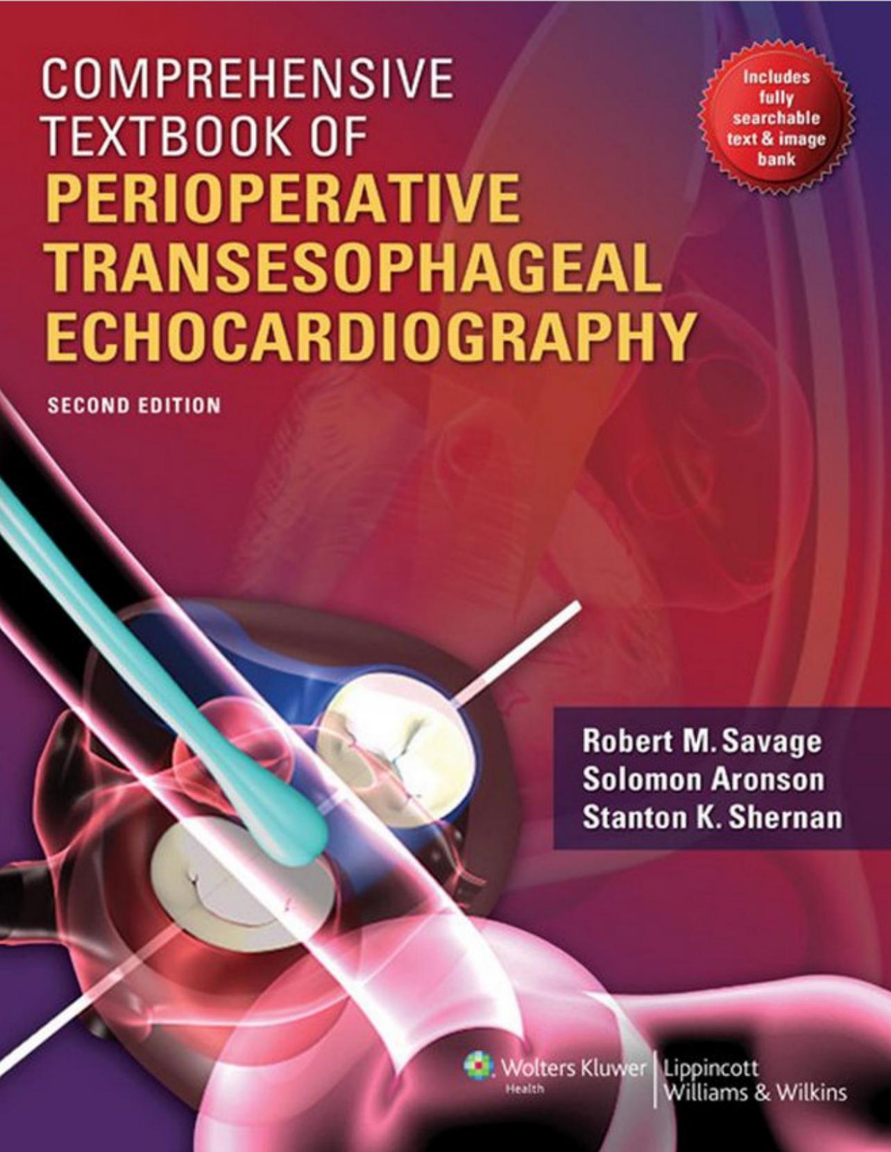
Comprehensive Textbook of Perioperative Transesophageal Echocardiography 2nd edition by Robert Savage,Solomon Aronson,Stanton Shernan 9781605472461 1605472468
$70.00 Original price was: $70.00.$35.00Current price is: $35.00.
Instant download Comprehensive Textbook of Perioperative Transesophageal Echocardiography Savage Robert M. Aronson Solomon Shernan Stanton K after payment
Comprehensive Textbook of Perioperative Transesophageal Echocardiography 2nd edition by Robert Savage,Solomon Aronson,Stanton Shernan – Ebook PDF Instant Download/Delivery:9781605472461,1605472468
Full dowload Comprehensive Textbook of Perioperative Transesophageal Echocardiography 2nd edition after payment
Product details:
ISBN 10:1605472468
ISBN 13: 9781605472461
Author:Robert Savage,Solomon Aronson,Stanton Shernan
Comprehensive Textbook of Perioperative Transesophageal Echocardiography 2nd Table of contents:
SECTION 1: Basic Perioperative Echocardiography
CHAPTER 1 Physics of Echocardiography
Introduction
Echocardiography and the Properties of Ultrasound
The Piezoelectric Effect and Ultrasound
Ultrasound Transducers: the Basics
Ultrasound Beam Geometry
Pulse-Echo Operation: Time Equals Distance
Interaction of Ultrasound with Tissues
Image Display Formats
2D Echocardiography: Transducer Operation, Frames, and Frame Rate
Tissue Harmonic Imaging
Contrast Echocardiography
Doppler Echocardiography
Continuous-Wave Doppler Echocardiography
Pulsed Doppler Echocardiography
Graphical Display of Doppler Frequency Spectra
Tissue Doppler Echocardiography
Color Flow Doppler Echocardiography
CW, PW, and Color Flow Doppler Compared
Conclusions
References
CHAPTER 2 Imaging Artifacts and Pitfalls
Artifacts
Missing Structures
Degraded Images
Falsely Perceived Objects
Misregistered Locations
Conclusion
References
CHAPTER 3 Optimizing Two-Dimensional Echocardiographic Imaging
Introduction
The Impact of Ultrasound Instrumentation on Image Generation and Display
Master Synchronizer (Clock)
Transducers and Spatial Resolution
Amplification and Time-Gain Compensation
Preprocessing
Analog-to-Digital Converters
Scan Conversion and Storage in Computer Memory
Postprocessing
Image Display, Recording, and Storage
Conclusions
References
CHAPTER 4 Surgical Anatomy of the Heart
Fibrous Skeleton of the Heart
Cardiac Ventricles
Tricuspid Valve
Pulmonic Valve
Mitral Valve Apparatus
Mitral Valve Leaflets: Carpentier-Sca Terminology
Mitral Valve Apparatus—Duran Terminology
Aortic Root
Coronary Anatomy
Conclusion
References
CHAPTER 5 Comprehensive and Abbreviated Intraoperative TEE Examination
Prevention of TEE Complications
Intraoperative TEE Indications
Optimizing Image Quality
Comprehensive TEE Examination
TEE Examination of the Thoracic Aorta
Abbreviated TEE Examination
TEE and Noncardiac Surgery
References
CHAPTER 6 Updated Indications for Intraoperative TEE
Practice Guidelines
Indications for Specific Lesions or Procedures
New Applications
Complications
References
CHAPTER 7 Organization of an Intraoperative Echocardiographic Service: Personnel, Equipment, Maintenance, Safety, Infection, Economics, and Continuous Quality Improvement
Personnel
Equipment and Maintenance
Safety
Economics of an Intraoperative Echo Service
Assessing Quality in an Intraoperative Tee Service
References
CHAPTER 8 Organizing Education and Training in Perioperative Transesophageal Echocardiography
Introduction
History of Training in TEE
Department of Cardiothoracic Anesthesia: Advanced Perioperative TEE Training
References
CHAPTER 9 Assessment of Global Ventricular Function
Normal Anatomy and Physiology of the Left Ventricle
Phases of Ventricular Systole
Left Ventricular Systolic Function
Two-Dimensional Examination of the Left Ventricle
Ejection Phase Indices of Left Ventricular Performance
Cardiac Output
Fractional Shortening
Fractional Area Change
Ejection Fraction
Calculating Preload and Afterload
Afterload
Systolic Index of Contractility (Dp/Dt)
Tissue Doppler Imaging
Speckle Tracking
Calculating Left Ventricular Filling Pressure
Interatrial Septum
Pulmonary Vein Flow
Mitral Inflow Velocities and Tissue Doppler
Three-Dimensional Evaluation of Global Ventricular Performance
References
CHAPTER 10 Assessment of Right Ventricular Function
Introduction
Structure and Function of the Right Ventricle
Transesophageal Echocardiographic Imaging of the Right Ventricle
Right Ventricular Hypertrophy and Dilatation
Right Ventricular Overload and the Ventricular Septum
Right Ventricular Systolic Function—Qualitative and Regional Assessment
Right Ventricular Global Systolic Function—Quantitative Echocardiographic Assessment
Right Ventricular Function—Hemodynamic Assessment
Right Ventricular Diastolic Function
References
CHAPTER 11 Assessment of Regional Ventricular Function
Introduction
Segmental Model of the Left Ventricle
Distribution of Coronary Artery Anatomy
Clinical Application of Regional Wall Motion Analysis
Assessment of Myocardial Viability
Emerging Echocardiographic Techniques for Assessing Regional Ventricular Function
References
CHAPTER 12 Assessment of Diastolic Dysfunction in the Perioperative Setting
Introduction
Clinical Importance of Diastolic Dysfunction
Physiology of Diastole and Pathophysiology of Dysfunction
Echocardiographic Evaluation of Left Ventricular Diastolic Function
Transmitral Inflow Velocities
Pulmonary Venous Flow
Mitral Annulus Tissue Doppler Imaging
Color M-Mode Doppler: Propagation Velocity
Grading of Diastolic Dysfunction
Limitations of Current Techniques of Echocardiographic Assessment of Diastolic Dysfunction
Emerging Techniques for Assessing LV Diastolic Function
Right Ventricular Diastolic Function
Clinical Implications of Diastolic Dysfunction in the Perioperative Setting
Conclusions
References
CHAPTER 13 Assessment of the Mitral Valve
Anatomy of the Mitral Valve
Structural Integrity of the Mitral Valve
ReferencesCHAPTER 14 Assessment of the Aortic Valve
Introduction
Anatomy
Approach
Aortic Stenosis
Aortic Regurgitation
References
CHAPTER 15 Assessment of the Tricuspid and Pulmonic Valves
Introduction
Structure and Function of the Tricuspid and Pulmonic Valves
Transesophageal Echocardiographic Evaluation of the Tricuspid and Pulmonic Valves
Congenital Diseases of the Tricuspid and Pulmonic Valves
Acquired Diseases of the Tricuspid and Pulmonic Valves
References
CHAPTER 16 Assessment of Prosthetic Valves
Types of Prosthetic Valves
Echocardiographic Evaluation of Prosthetic Heart Valves
Normal Imaging
Hemodynamic Measurements
Echocardiographic Characteristics of Specific Valve Types
Prosthetic Valve Dysfunction and Complications
Associated Complications
References
CHAPTER 17 Assessment of Cardiac Masses
Approach/Structures
Ivc/Svc/Right Atrium
Right Ventricle/Pulmonary Artery
Pulmonary Veins/Left Atrium/Left Atrial Appendage
Left Ventricle
Aorta/Valves
Pathology
Ivc/Svc/Right Atrium
Right Ventricle/Pulmonary Artery
Pulmonary Veins/Left Atrium/Left Atrial Appendage
Left Ventricle
Aorta/Valves
References
CHAPTER 18 Transesophageal Echocardiographic Evaluation for Noncardiac Surgery
Tee as a Diagnostic Rescue Tool
Tee as a Tool for Monitoring Cardiac Performance
Tee as a Confirmation Tool
Conclusions
References
CHAPTER 19 Ultrasound for Vascular Access
Indications for Vascular Access
Technical Considerations
Ultrasound-Guided Central Venous Access
Ultrasound-Guided Arterial Access
Development of Ultrasound Vascular Access Service
Future Directions
References
SECTION 2: Echocardiography in the Critical Care Setting
CHAPTER 20 Overview and Relevance of Transesophageal Echocardiography in Critical Care Medicine
Imaging Orientation and Nomenclature
Indications and Contraindications
Effect and Importance of Echocardiography
References
CHAPTER 21 Chest Wall Echocardiography in the Intensive Care Unit
Introduction
Basic Imaging Using Transthoracic Echocardiography
Hemodynamic Assessment of the Critically ill Patient
Valvular Function and Disease
Pulmonary Embolism
Pericardial Disease
Myocardial Ischemia and Its Complications
Intracardiac and Intrapulmonary Shunts
Conclusion
References
CHAPTER 22 The Assessment of a Patient with Endocarditis
Introduction
Epidemiology and Risk Factors
Diagnosis
Echocardiography
Surgical Indications for Infective Endocarditis
Intraoperative Assessment
Conclusion
References
CHAPTER 23 Rescue Echocardiography in the Critically Ill Patient
Unrecognized Hypovolemia
Pulmonary Embolism
Ventricular Dysfunction
Aortic Dissection
Severe Valvular Disease
Intracardiac Thrombi
Ischemia Monitoring
Pericardial Fluid
References
SECTION 3: Advanced Applications in Perioperative Echocardiography
CHAPTER 24 Epiaortic and Epicardial Imaging
Epicardial Imaging
Epiaortic Imaging
Technique
References
CHAPTER 25 Assessment of Congenital Heart Disease in the Adult Patient
Indications
Simple Lesions
Complex Lesions
Conclusions
References
CHAPTER 26 Assessment of Perioperative Hemodynamics
Doppler Measurements of Stroke Volume and Cardiac Output
Doppler Measurement of Pulmonary-To-Systemic Flow Ratio (Qp/Qs) (Fig. 26.12)
Doppler Assessment of Regurgitation
Doppler Measurement of Pressure Gradients
Doppler Determination of Valve Area
Doppler Determination of Intracardiac Pressures
Doppler Measurement of Dp/Dt
References
CHAPTER 27 Assessment in Higher Risk Myocardial Revascularization and Complications of Ischemic Heart Disease
Historical Perspectives
Indications for Myocardial Revascularization
Definition of Higher Risks in Patients Undergoing Coronary Artery Bypass Graft Surgery
Importance of Ischemic Heart Disease in Future Health Care Delivery
Principles of Performing Intraoperative Echo Examination in Cabg Surgery
Complications of Ischemic Heart Disease
Future Applications Table 27.14
References
CHAPTER 28 Assessment of the Mitral Valve in Ischemic Heart Disease
Scientific Principles
The Echocardiographic Examination
Procedure Evaluation
References
CHAPTER 29 Surgical Considerations in Mitral and Tricuspid Valve Surgery
Primary Mitral Valve Disease
Prosthetic Mitral Disease
Tricuspid Valve
Structure and Anatomy
Pathology
Surgical Management of Mitral Valve Disease
Surgical Procedures for Mitral Valve Regurgitation
Ring Annuloplasty
Triangular Resection and Annuloplasty Ring for Posterior Leaflet Prolapse or Flail
Chordal Transfer for Ruptured or Elongated Segment of the
Surgical Procedures for Mitral Valve Stenosis
Surgical Management of Tricuspid Valve Disease
Tricuspid Valve Replacement
References
CHAPTER 30 Surgical Considerations and Assessment in Nonischemic Mitral Valve Surgery
Historical Perspectives
Importance of Intraoperative Echocardiography (IOE) in Mv Surgery
Importance of Intraoperative Echocardiograpy (IOE) Mv Assessment in the Future of Cardiothoracic Anesthesia
Intraoperative Echocardiography and Critical Issues in MV Surgery
Organization of Chapter
Key Concepts
Mitral Valve Apparatus
Correlation With Imaging Planes: the 2D Examination of the Mitral Valve
Pathology
Mitral Valve Procedures
References
CHAPTER 31 Assessment in Aortic Valve Surgery
Critical Issues During Aortic Valve Surgery
Role of TEE in Surgical Decision Making
Approach
Specific Aortic Valve Procedures
Conclusions
References
CHAPTER 32 Surgical Considerations and Assessment in Aortic Surgery
Introduction
Anatomy of the Thoracic Aorta
Overview of Thoracic Aortic Diseases
Transesophageal Echocardiography in the Examination of the Aorta
Intraoperative Role of TEE in Aortic Disease
Conclusions
References
CHAPTER 33 Surgical Considerations and Assessment of Endovascular Management of Thoracic Vascular Disease
Introduction
Anatomy of the Thoracic Aorta
Thoracic Aortic Aneurysms
Thoracic Aortic Dissections
Thoracic Aortic Transections
Other Aortic Pathologies
Current Practice of Endovascular Repair of the Thoracic Aorta
Assessment of Thoracic Aortic Pathologies
Role of Transesophageal Echocardiography
Role of Intravascular Ultrasound
References
CHAPTER 34 Echocardiographic Assessment of Cardiomyopathies
Introduction
Dilated Cardiomyopathy
Hypertrophic Cardiomyopathy
Restrictive Cardiomyopathy
Arrhythmogenic Right Ventricular Cardiomyopathy
Unclassified Cardiomyopathies
Role of Echocardiography: Prognostication and Treatment
References
CHAPTER 35 Surgical Considerations and Assessment in Heart Failure Surgery
Impact of Congestive Heart Failure on the Population
Trends in Management of Patients with Congestive Heart Failure
Critical Issues Addressed by Intraoperative Echocardiography: Patients Undergoing Procedures for CHF
Intraoperative Echocardiography in Surgical Procedures for CHF
Ventricular Constraint Techniques
Mechanical Circulatory Support
Conclusion
References
CHAPTER 36 Assessment of Cardiac Transplantation
The Role of TEE in Cardiac Donor Screening
Intraoperative Monitoring in the Pretransplantation Period
Management of Early Postoperative Hemodynamic Abnormalities in the Intensive Care Unit
Postoperative Follow-up Studies of Cardiac Allograft Function
References
CHAPTER 37 Assessment in Cardiac Intervention
Percutaneous Aortic Valve Replacement
Mitral Valve Repair
Nonsurgical Septal Reduction for Hypertrophic Cardiomyopathy
Pericardiocentesis for Cardiac Tamponade
Myocardial Biopsy
Mitral Balloon Valvulotomy
Radiofrequency Ablation
Conclusion
References
CHAPTER 38 Perioperative Application of New Modalities: Strain Echocardiography and Three-Dimensional Echocardiography
Introduction
Myocardial Structure and Motion
Deformation
Principles of Doppler Tissue Imaging and Doppler Strain Echocardiography
Step-By-Step Guide on How to Obtain Doppler Strain
Principles of Speckle Tracking Imaging and 2d Strain
Shear Strain and Torsion
Step-By-Step Guide on How to Perform STI
Correlation of Strain Between Dti and STI
Applications of DTI- and STI-Derived Strain
Three-Dimensional Echocardiography
References
CHAPTER 39 Echocardiographic Evaluation of Pericardial Disease
Pericardial Anatomy
Pericardial Physiology
Pericardial Pathology
People also search for Comprehensive Textbook of Perioperative Transesophageal Echocardiography 2nd:
perioperative transesophageal echocardiography
a comprehensive guide to intersex
comprehensive tee guidelines
transesophageal echocardiography books
comprehensive tee exam
comprehensive textbook of suicidology


