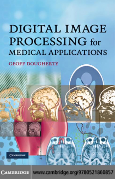
Digital Image Processing for Medical Applications 1st edition by GEOFF DOUGHERTY ISBN 0511530374 9780511530371
$70.00 Original price was: $70.00.$35.00Current price is: $35.00.
Instant download Digital Image Processing for Medical Applications GEOFF DOUGHERTY after payment
Digital Image Processing for Medical Applications 1st edition by GEOFF DOUGHERTY – Ebook PDF Instant Download/Delivery: 0511530374 , 9780511530371
Full download Digital Image Processing for Medical Applications 1st edition after payment

Product details:
ISBN 10: 0511530374
ISBN 13: 9780511530371
Author: GEOFF DOUGHERTY
Image processing is a hands-on discipline, and the best way to learn is by doing. This text takes its motivation from medical applications and uses real medical images and situations to illustrate and clarify concepts and to build intuition, insight and understanding. Designed for advanced undergraduates and graduate students who will become end-users of digital image processing, it covers the basics of the major clinical imaging modalities, explaining how the images are produced and acquired. It then presents the standard image processing operations, focusing on practical issues and problem solving. Crucially, the book explains when and why particular operations are done, and practical computer-based activities show how these operations affect real images. All images, links to the public-domain software ImageJ and custom plug-ins, and selected solutions are available from www.cambridge.org/books/dougherty.
Digital Image Processing for Medical Applications 1st Table of contents:
Part I: Introduction to Image Processing
1. Introduction
-
Overview
-
Learning objectives
1.1 Imaging systems
1.2 Objects and images
1.3 The digital image processing system
1.4 Applications of digital image processing -
Exercises
2. Imaging Systems
-
Overview
-
Learning objectives
2.1 The human visual pathway-
2.1.1 Brightness response of the eye
-
2.1.2 Spatial resolution of the eye
2.2 Photographic film -
2.2.1 Response of film to light
-
2.2.2 Spatial resolution of film
2.3 Other sensors
2.4 Digitizing an image -
2.4.1 Spatial quantization
-
2.4.2 Intensity quantization
2.5 The quality of a digital image -
2.5.1 Spatial resolution and pixel size
-
2.5.2 Brightness resolution
-
2.5.3 Noise content
2.6 Color images
-
-
Computer-based activities
-
Exercises
3. Medical Images Obtained with Ionizing Radiation
-
Overview
-
Learning objectives
3.1 Medical imaging modalities
3.2 Images from x-rays-
3.2.1 Plain (film-screen) radiography
-
Unsharpness
-
Contrast
-
-
3.2.2 Computed radiography
-
3.2.3 Mammography
-
3.2.4 Fluoroscopy and digital subtraction angiography
-
3.2.5 Computed tomography
3.3 Images from γ-rays -
3.3.1 Planar scintigraphy
-
3.3.2 SPECT imaging
-
3.3.3 PET imaging
3.4 Dose and risk
-
-
Computer-based activities
-
Exercises
4. Medical Images Obtained with Non-Ionizing Radiation
-
Overview
-
Learning objectives
4.1 Ultrasound imaging-
4.1.1 Image quality
-
4.1.2 Doppler imaging
-
4.1.3 Clinical applications of ultrasound
4.2 Magnetic resonance imaging -
4.2.1 Nuclear magnetic resonance
-
4.2.2 Magnetic resonance imaging (MRI)
-
4.2.3 Pulse sequences
-
4.2.4 T1- and T2-weighted images
-
4.2.5 Image characteristics
4.3 Picture archiving and communication systems (PACS) -
4.3.1 Multimodal registration
-
-
Computer-based activities
-
Exercises
Part II: Fundamental Concepts of Image Processing
5. Fundamentals of Digital Image Processing
-
Overview
-
Learning objectives
5.1 The gray-level histogram-
5.1.1 Dynamic range and contrast
-
5.1.2 Entropy
-
5.1.3 Signal-to-noise ratio
-
5.1.4 Other histogram features
5.2 Histogram transformations and look-up tables -
5.2.1 Histogram stretch
-
5.2.2 Histogram equalization
-
5.2.3 Histogram matching
-
5.2.4 Local histogram transformations
-
5.2.5 Other histogram transformations
-
-
Computer-based activities
-
Exercises
6. Image Enhancement in the Spatial Domain
-
Overview
-
Learning objectives
6.1 Algebraic operations-
6.1.1 Averaging
-
6.1.2 Image subtraction
-
6.1.3 Multiplication and division
6.2 Logical (Boolean) operations
6.3 Geometric operations -
6.3.1 The log-polar transformation
6.4 Convolution-based operations -
6.4.1 Smoothing masks
-
6.4.2 Sharpening and edge-detecting masks
-
6.4.3 Second-derivative masks
-
-
Computer-based activities
-
Exercises
7. Image Enhancement in the Frequency Domain
-
Overview
-
Learning objectives
7.1 The Fourier domain
7.2 The Fourier transform
7.3 Properties of the Fourier transform
7.4 Sampling-
7.4.1 Window functions
-
7.4.2 Aliasing
-
7.4.3 Sub-sampling
-
7.4.4 Reconstruction from samples
7.5 Cross-correlation and autocorrelation
7.6 Imaging systems point spread function and optical transfer function
7.7 Frequency domain filters -
7.7.1 Low-pass or smoothing filters
-
7.7.2 High-pass or sharpening filters
-
7.7.3 Band-pass and band-reject filters
-
7.7.4 Homomorphic filters
-
7.7.5 Spatial masks vs. frequency filters
7.8 Tomographic reconstruction -
7.8.1 Backprojection
-
7.8.2 Direct Fourier reconstruction
-
-
Computer-based activities
-
Exercises
8. Image Restoration
-
Overview
-
Learning objectives
8.1 Image degradation
8.2 Noise-
8.2.1 Types of noise
-
White noise
-
Colored noise
-
Periodic noise
-
Gaussian noise
-
Impulse (or salt-and-pepper) noise
-
Quantization noise
-
Photon noise (also called quantum noise or shot noise)
-
Speckle (or multiplicative) noise
8.3 Noise-reduction filters
-
-
8.3.1 Adaptive filters
8.4 Blurring -
8.4.1 Deblurring
8.5 Modeling image degradation -
8.5.1 Wiener filters
-
8.5.2 Other filters
-
8.5.3 Blind image restoration
8.6 Geometric degradations
-
-
Computer-based activities
-
Exercises
Part III: Image Analysis
9. Morphological Image Processing
-
Overview
-
Learning objectives
9.1 Mathematical morphology-
9.1.1 Connectivity
9.2 Morphological operators -
9.2.1 Dilation and erosion
-
9.2.2 Opening and closing
-
9.2.3 Hit-or-miss transform
-
9.2.4 Thinning and skeletonization
-
9.2.5 The medial axis transform and skeletonization
-
9.2.6 The convex hull
9.3 Extension to grayscale images -
9.3.1 Applications of grayscale morphological processing
-
-
Computer-based activities
-
Exercises
10. Image Segmentation
-
Overview
-
Learning objectives
10.1 What is segmentation?
10.2 Thresholding-
10.2.1 Optimal thresholding
-
10.2.2 Adaptive thresholding
10.3 Region-based methods
10.4 Boundary-based methods -
10.4.1 Edge detection and linking
-
10.4.2 Boundary tracking
10.5 Other methods -
10.5.1 Active contours
-
10.5.2 Watershed segmentation
-
-
Computer-based activities
-
Exercises
11. Feature Recognition and Classification
-
Overview
-
Learning objectives
11.1 Object recognition and classification
11.2 Connected components labeling
11.3 Features
11.4 Object recognition and classification-
11.4.1 Classification
11.5 Statistical classification -
11.5.1 Parametric methods
-
11.5.2 Non-parametric techniques
-
11.5.3 Unsupervised methods
-
k-means clustering
-
Hierarchical clustering
11.6 Structural/syntactic classification
11.7 Applications in medical image analysis
-
-
-
Computer-based activities
-
Exercises
12. Three-Dimensional Visualization
-
Overview
-
Learning objectives
12.1 Image visualization
12.2 Surface rendering
12.3 Volume rendering
12.4 Virtual reality -
Computer-based activities
-
Exercises
Part IV: Medical Applications and Ongoing Developments
13. Medical Applications of Imaging
-
Overview
-
Learning objectives
13.1 Computer-aided diagnosis in mammography
13.2 Tumor imaging and treatment
13.3 Angiography
13.4 Bone strength and osteoporosis
13.5 Tortuosity
14. Frontiers of Image Processing in Medicine
-
Overview
-
Learning objectives
14.1 Trends-
14.1.1 The inverse problem
-
14.1.2 Functional magnetic resonance imaging (fMRI)
-
14.1.3 Molecular imaging
-
14.1.4 Other imaging modalities
-
14.1.5 Surgical interventions
14.2 The last word
-
Appendix A: The Fourier Series and Fourier Transform
Appendix B: Set Theory and Probability
-
B.1 Concepts from set theory
-
B.2 Boolean algebra/logic operations
-
B.3 Probability
-
B.4 Image segmentation
-
Computer-based activities
-
Exercises
Appendix C: Shape and Texture
-
C.1 Shape
-
C.2 Fractals
-
C.3 Texture
-
C.3.1 Statistical approaches
-
C.3.2 Structural approaches
-
C.3.3 Spectral approaches
-
People also search for Digital Image Processing for Medical Applications 1st :
what is used in medical imaging
digital image processing for medical applications pdf
digital image processing applications in medical field
digital image processing in medical field
applications of digital image processing in medical field
Tags: GEOFF DOUGHERTY, Digital Image, Medical Applications


